Videos
Above and below are links to videos associated with published papers from the lab. The videos show myoclonic twitching during sleep in infant rodents and humans; some videos also show associated brain activity. All links to videos take you to a new page on the host site, Databrary.
-
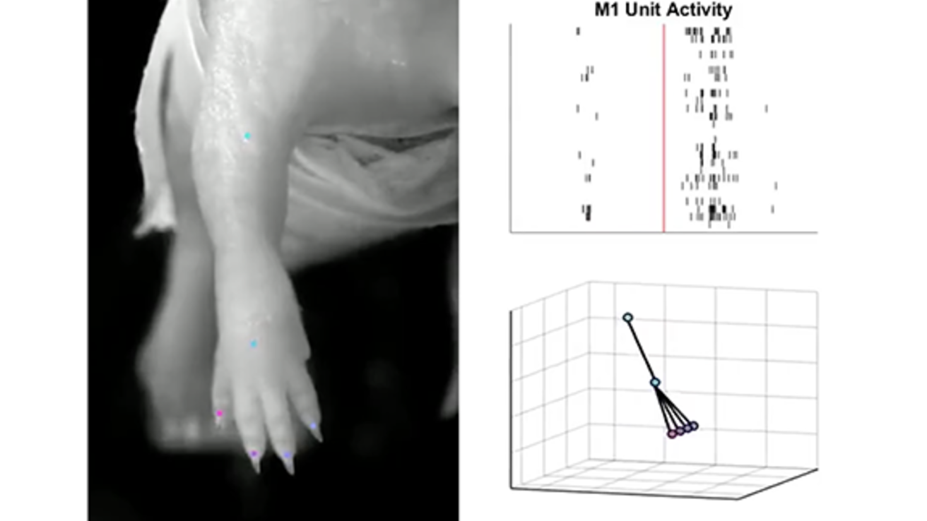
Movie 1 and Movie 2 from Glanz et al., Sensory coding of limb kinematics in motor cortex across a key developmental transition. Journal of Neuroscience, 41: 6905-6918, 2021.
-
Movie 1: Relationship between forelimb twitching (left) and unit activity in the contralat- eral forelimb region of M1 (top right) in a P8 rat. Limb movement was tracked using DeepLabCut and visualized in MATLAB (bottom right). Each row in the M1 record represents 1 of 26 simultaneously recorded units; each unit is associated with a distinct audible tone. Note the discontinuous pattern of M1 activity at this age.
-
Movie 2: Same as for Movie 1 except for 31 M1 units in a P12 rat. Note the continuous pattern of M1 activity at this age.
-
-
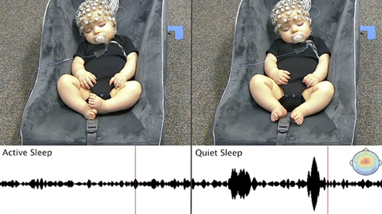
Video S1 from Sokoloff et al., Twitches emerge postnatally during quiet sleep in human infants and are synchronized with sleep spindles. Current Biology, 31: 3426-3432, 2021.
-
Twitching during AS (left) and QS (right) in a 6-month-old human infant, related to Figure 1 in the paper. Most of the twitches occur in the hands, fingers, feet, and toes. Each recording is 30 s in duration and playback speed is at 3x. Two sleep spindles are shown during QS, recorded at electrode C3 and bandpass filtered (12-14 Hz).
-
-
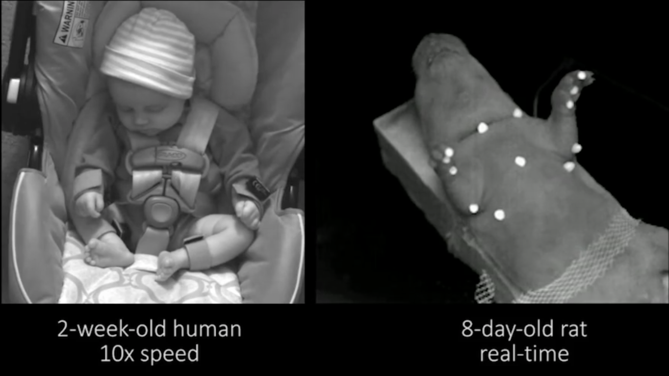
Video S1 and Video S2 from Sokoloff et al.. Spatiotemporal organization of myoclonic twitching in sleeping human infants. Developmental Psychobiology, 62: 697-710, 2020.
-
Video S1: Three clips showing twitching in a 2-week-old human infant during active (REM) sleep. The clips are played back at three speeds: normal, 3x, and 10x. Twitches can be seen in the arms, legs, head, and face. At the end of the third clip, the baby wakes up (head, arms, and legs lift and flail).
-
Video S2: Comparison of twitching between a 2-week-old human infant at 10x normal playback speed and a P8 rat at normal playback speed.
-
-
Movie S1 from Blumberg et al., Development of twitching in sleeping infant mice depends on sensory experience. Current Biology, 25:656-662, 2015.
-
In a P10 mouse, a twitch pair comprising a shoulder abduction followed by a shoulder adduction is shown in two successive clips at two speeds: at 1/40th real time and at 1/4th real time. As shown, the left forelimb begins at a resting position, exhibits a shoulder abduction followed immediately by a shoulder adduction, and then returns to the resting position.
-
-
Movie S1 from Tiriac et al.. Self-generated movements with "unexpected" sensory consequences. Current Biology, 24:2136-2141, 2014.
-
In these clips of a P10 rat, the left hindlimb of the P10 rat is closest to the viewer and the multiunit activity, depicted at the bottom, was recorded from the contralateral (i.e., right) M1. Eight clips are shown: (i) manual stimulation of the hindlimb contralateral to M1; (ii) manual stimulation of the hindlimb ipsilateral to M1; (iii) spontaneous myoclonic twitching during active sleep in real-time; (iv) spontaneous myoclonic twitching during active sleep in slow motion; (v) spontaneous wake movements; (vi) hindlimb movements evoked by systemic administration of quipazine; (vii) hindlimb movements evoked by generalized arousal produced by application of a cold stimulus to the pup's snout; and (viii) hindlimb movements evoked reflexively by flicking the tail. All videos were recorded at 30 frames per second.
-
-
Movie S1 from Blumberg et al.. Spatiotemporal structure of REM sleep twitching reveals developmental origins of motor synergies. Current Biology, 23:2100-2013.
-
In a P8 rat, five clips are shown: (i) 18 seconds of real-time twitching; (ii) a discrete twitch, in slow motion, comprising extension of the left elbow; (iii) a discrete twitch, in slow motion, comprising abduction of the right shoulder; (iv) an example, in slow motion, of a homologous twitch pattern comprising right shoulder adduction followed quickly by left shoulder adduction; and (v) an example, in slow motion, of a complex multi-joint twitch pattern comprising several movements across both forelimbs. The white dots are fluorescent paint for motion tracking of joint movements. All videos were recorded at 250 frames per second.
-
-
Movie S1 from Tiriac et al.. Rapid whisker movements in sleeping newborn rats. Current Biology, 22:2075-2080, 2012.
-
Sample video clips of individual, dual, and multiple whisker twitches in P4 rats are shown, as is an example of a mystacial pad movement. See Figure 1A in the paper to orient the whiskers on the snout and to identify each of the 11 whiskers and Figure 1B for the associated quiver plots. Each clip was recorded at 200 frames/s and is played back at 50 frames/s. The white light in the lower-right-hand corner indicates that the experimenter observed twitching of the distal limbs or tail, indicative of active sleep. Note that these clips only show those 11 whiskers that were monitored in these tests.
-